20 November 2006, Volume 45, Issue 33, pp. 8393-8565
24 articles
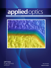
Feasibility of tomographic oxygen imaging by phosphorescence lifetime was proven by both numerical simulations and phantom experiments, using newly developed dendritically protected near-infared oxygen probes (top). Volumetric lifetime distributions were obtained by fitting a series of independently reconstructed images of integrated phosphorescence photon density, taking advantage of large differences between the time scales of photon migration and phosphorescence emission (bottom). By varying the length of excitation pulses and delaying the integration of signal collection, the contrast in images of hypoxic objects could be brought up to several hundreds. For details, see the paper by Apreleva et al., pp. 8547-8559.